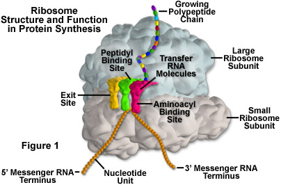
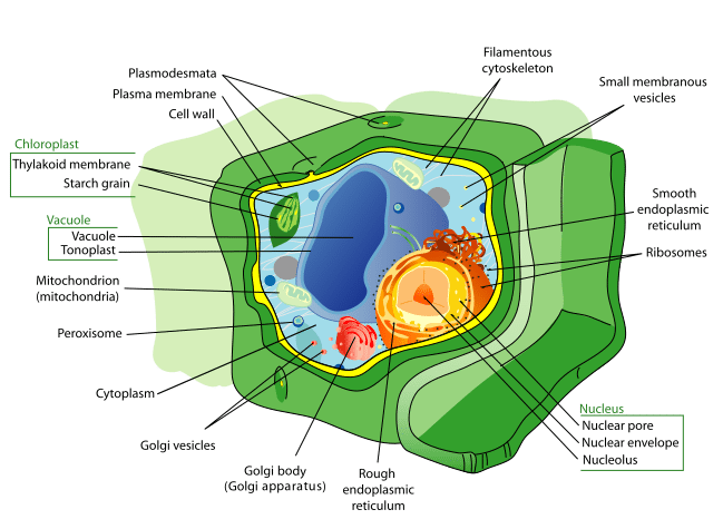
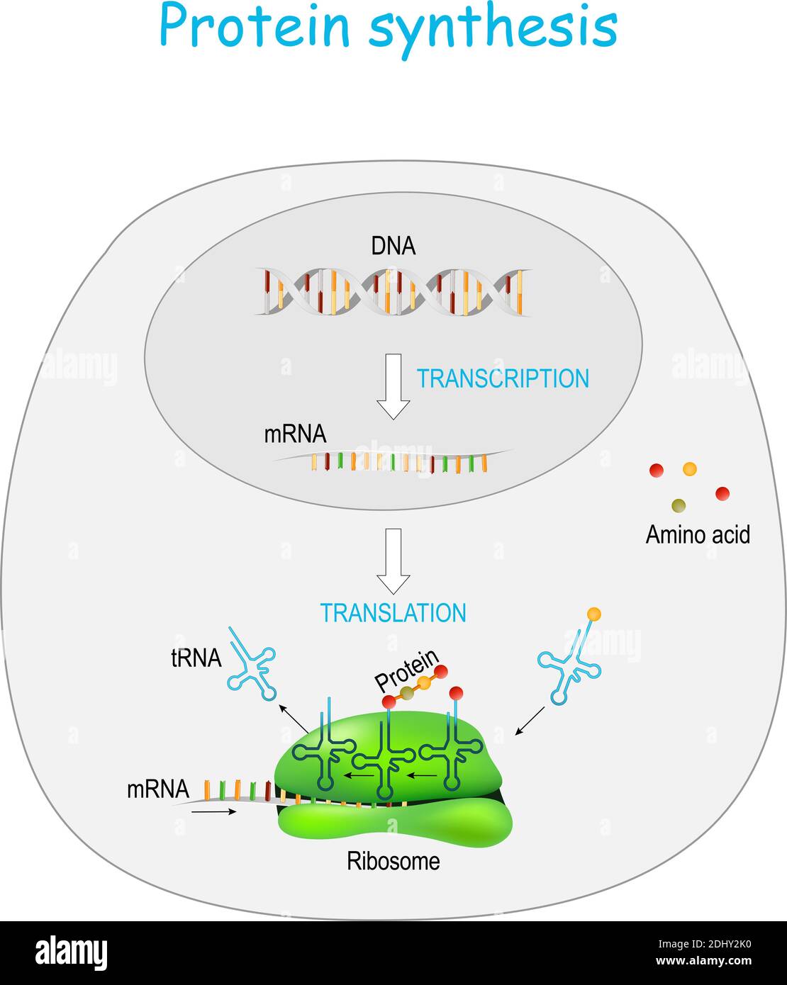
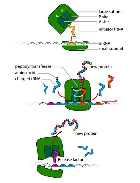
41 ribosome diagram with labels
quizlet.com › 520218106 › science-s1-flash-cardsScience S1 Flashcards | Quizlet Study with Quizlet and memorize flashcards containing terms like In which order did the events forming our solar system occur? The solar nebula became hot and dense pulling in more gas.This flattened into a rotating disk. It spun faster and faster, forming the Sun. Gas was pulled toward the center, forming the Sun. Gas flattened into a rotating disk and became hot and dense, forming a solar ... Ribosome - Wikipedia During 1977, Czernilofsky published research that used affinity labeling to identify tRNA-binding sites on rat liver ribosomes. Several proteins, including L32/33, L36, L21, L23, L28/29 and L13 were implicated as being at or near the peptidyl transferase center. [27] Plastoribosomes and mitoribosomes [ edit]
ER proteins decipher the tubulin code to regulate organelle 15/12/2021 · Peak labels show number of glutamates on α- and β-tubulin. Glutamate numbers are indicated in green, dark green, orange, and red for α1B, α1C, βI and βIVb isoforms, respectively.

Ribosome diagram with labels
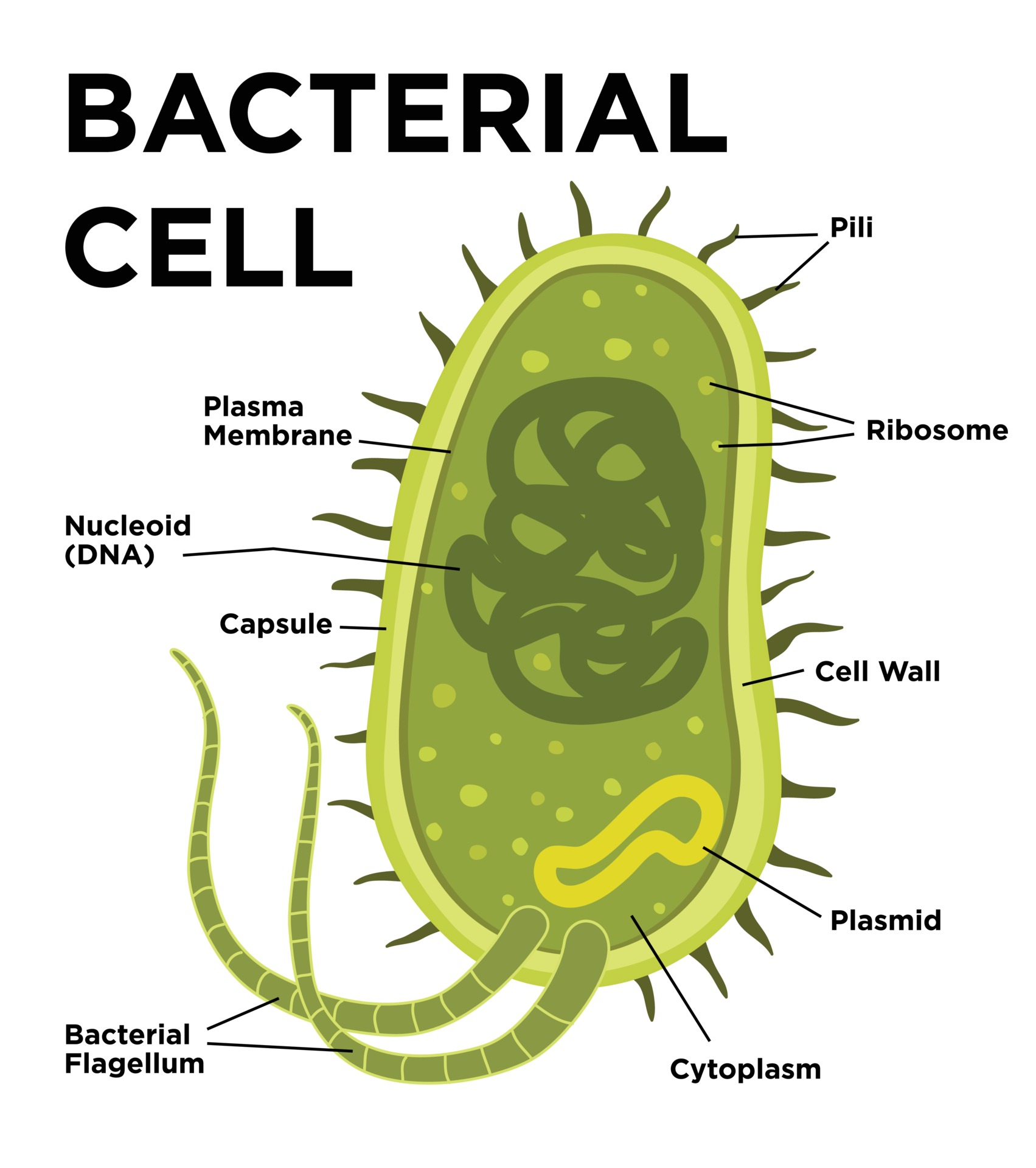
Ribosome - Definition, Function and Structure | Biology Dictionary A. Ribosomes translate the 4 base language of DNA into the 20 base language of proteins, allowing for many more combinations. B. The 4 different nucleobases of DNA can be recombined endlessly to produce new proteins. C. Ribosomes can modify proteins with carbohydrates to make them unique. Answer to Question #2 3. Animal Cell Diagram | Science Trends The diagram, like the one above, will include labels of the major parts of an animal cell including the cell membrane, nucleus, ribosomes, mitochondria, vesicles, and cytosol. The cells of animals are the basic structural units for the wide variety of life we see in the animal kingdom. Structure of Ribosome (With Diagram) - Biology Discussion A bacterial ribosome is about 250 nm in diameter and consists of two subunits, one large and one small. Both subunits consist of one or more molecules of rRNA and an array of ribosomal proteins. ADVERTISEMENTS: Association of two subunits is called mono-some. The structure of prokaryotic ribosome is given in the figure 8.2 B.
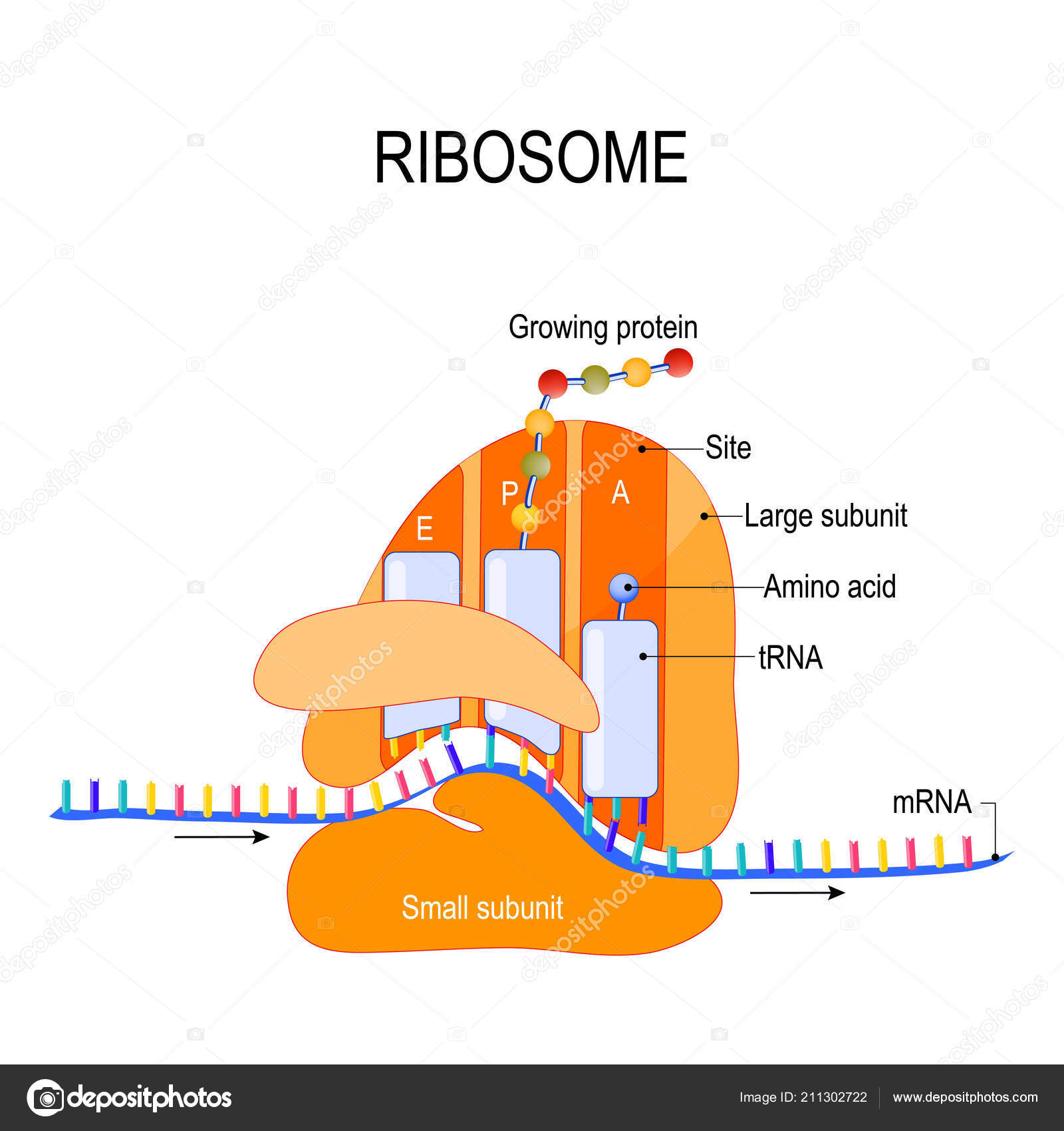
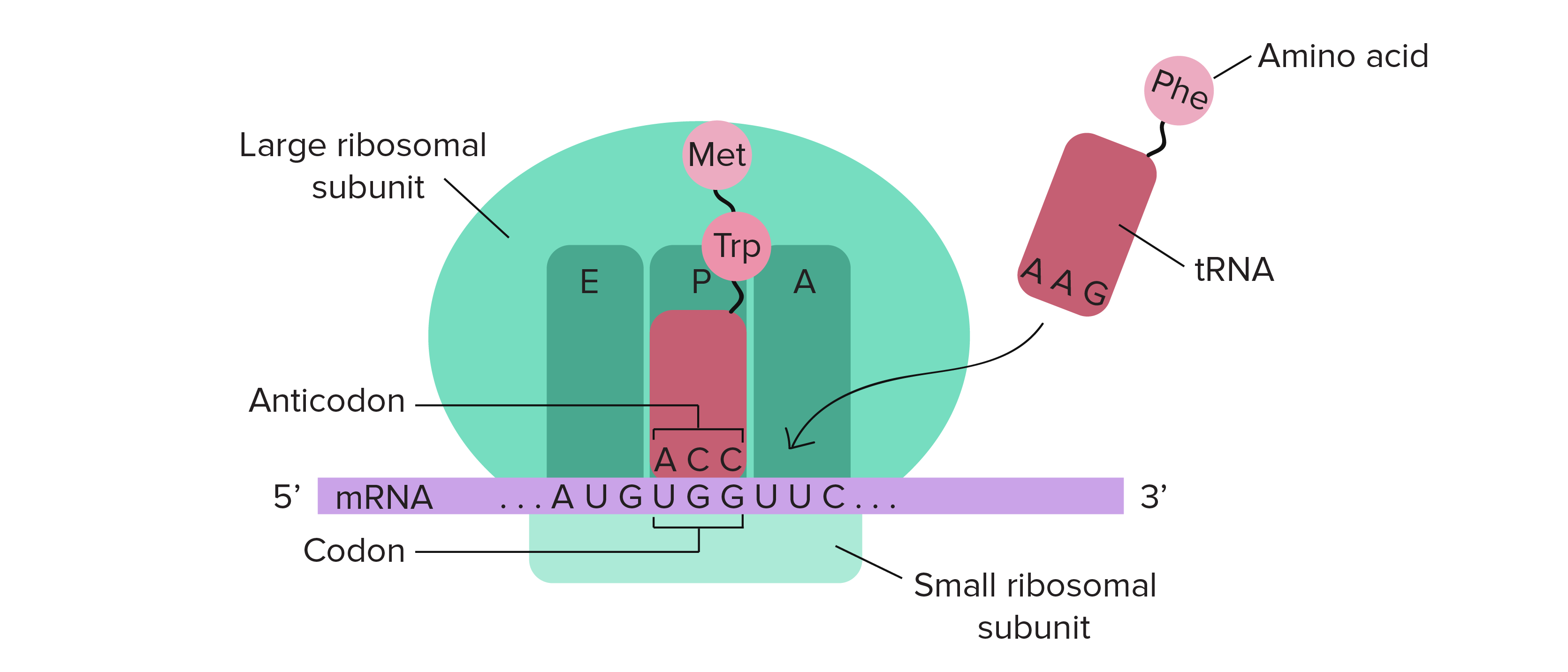
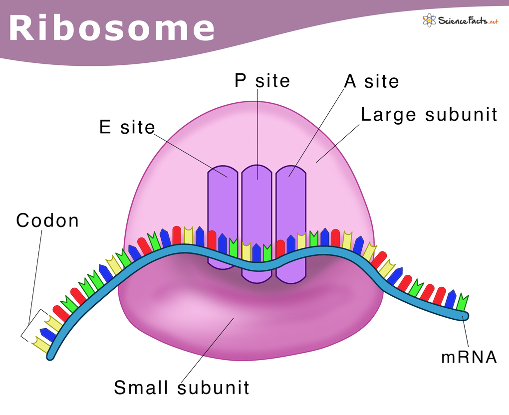
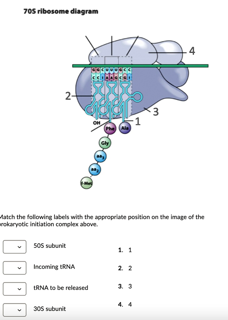
Ribosome diagram with labels. en.wikipedia.org › wiki › Nuclear_envelopeNuclear envelope - Wikipedia The nuclear envelope is punctured by around a thousand nuclear pore complexes, about 100 nm across, with an inner channel about 40 nm wide. The complexes contain a number of nucleoporins, proteins that link the inner and outer nuclear membranes. AAMC MCAT Practice Exam 4 Bb Solutions - MCAT Content 21) When assembled for translation, ribosomes have three binding sites that accommodate tRNAs: The A site, the P site, and the E site. Take a look at the diagram below to see how these are arranged relative to each other: Incoming aminoacyl-tRNAs (a tRNA with an amino acid covalently attached) enter the ribosome at the A site. The peptidyl-tRNA ... Solved In the following diagram of a ribosome, assign the - Chegg In the following diagram of a ribosome, assign the correct labels. Who are the experts? Experts are tested by Chegg as specialists in their subject area. We review their content and use your feedback to keep the quality high. Transcribed image text: In the following diagram of a ribosome, assign the correct labels. DNA Labeling: Transciption and Translation - The Biology Corner This worksheet shows a diagram of transcription and translation and asks students to label it; also includes questions about the processes. Name: _____ ... How does the ribosome know the sequence of amino acids to build? 12. What is the difference between a codon and an anticodon?
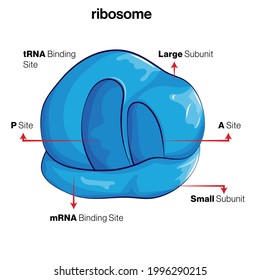
Ribosomes Images Stock Photos, Pictures & Royalty-Free Images - iStock Ribosomes are present in the cells of all forms of life, from bacteria to humans. DNA is copied to RNA, and Ribosomes read the instructions encoded in RNA to build proteins. Ribosomes were first observed in the 1950s, but the detail of their complex structure wasn't known until the early 2000s. Vector diagram of Mitochondria. Cross-section view. Answered: a) What is a mutation in molecular… | bartleby Add labels to the strands above to show the 3’ and 5’ ends. arrow_forward. Using the following list of codons describe, using diagrams etc., how information stored in the DNA is translated into a peptide. Be sure to discuss all steps. In other words, use a diagram and give me sequences, transcription and translation steps. Show the sequences of the sense and the other DNA … Mastering Microbiology Chapter 7 Flashcards | Quizlet Ribosomes use mRNA as instructions, which provide a code specifying the order of amino acids in a protein. ... Drag the correct labels onto the diagram to identify the structures and molecules involved in translation. a. mRNA b. small subunit of ribosome c. large subunit of ribosome Ribosomes- Definition, Structure, Functions and Diagram - Microbe Notes Ribosomes Definition The ribosome word is derived - 'ribo' from ribonucleic acid and 'somes' from the Greek word 'soma' which means 'body'. Ribosomes are tiny spheroidal dense particles (of 150 to 200 A0 diameters) that are primarily found in most prokaryotic and eukaryotic. They are sites of protein synthesis.
Cell Organelles- Definition, Structure, Functions, Diagram Animal Cell- Definition, Structure, Parts, Functions, Labeled Diagram; Prokaryotes vs Eukaryotes- Definition, 47 Differences, Structure, Examples; Amazing 27 Things Under The Microscope With Diagrams ... Ribosomes. Ribosomes are ribonucleoproteins containing equal parts RNA and proteins along with an array of other essential components required ... Labeled Plant Cell With Diagrams | Science Trends The ribosomes are created in the nucleolus of the cell. Ribosomes are made out of two smaller subunits, a large ribosomes subunit and a small ribosomal subunits. The transfer RNA or tRNA encodes the correct series of genetic instructions into the mRNA or messenger RNA, which is what ensures that the right proteins are created. Ribosomes Illustrations & Vectors - Dreamstime Download 657 Ribosomes Stock Illustrations, Vectors & Clipart for FREE or amazingly low rates! New users enjoy 60% OFF. 191,573,101 stock photos online. ... Anatomical and medical labeled scheme. Explained closeup diagram. Ribosomes vector illustration. Anatomical and medical labeled. Ribosome - protein factory - definition, function, structure and biology The protein translation by a ribosome consists of three stages: (1) Initiation, (2) Elongation, and (3) Termination. Initiation - the ribosome assembles around the target mRNA. A small ribosome subunit links onto the "start-end" of an mRNA strand. "Initiator tRNA" also enters the small subunit and binds to the start codon (most commonly, AUG).
Animal Cells: Labelled Diagram, Definitions, and Structure - Research Tweet Ribosomes Ribosomes create proteins. They can float freely in the cytoplasm or can be attached to the nuclear envelope. They create proteins by assembling amino acids into polypeptides. As the ribosomes build an amino acid chain, the chain is pushed into the endoplasmic reticulum.
Science S1 Flashcards | Quizlet Study with Quizlet and memorize flashcards containing terms like In which order did the events forming our solar system occur? The solar nebula became hot and dense pulling in more gas.This flattened into a rotating disk. It spun faster and faster, forming the Sun. Gas was pulled toward the center, forming the Sun. Gas flattened into a rotating disk and became hot and dense, forming …
en.wikipedia.org › wiki › Biuret_testBiuret test - Wikipedia The Biuret (IPA: / ˌ b aɪ j ə ˈ r ɛ t /, / ˈ b aɪ j ə ˌ r ɛ t /) test, also known as Piotrowski's test, is a chemical test used for detecting the presence of peptide bonds (Note that at least two peptide bonds are needed to be present in the molecule to show this test).
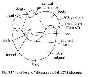
Ribosome Images - University of California, Santa Cruz 70S Ribosome (left side view) with labels. JPG | TIFF 70S Ribosome (30S view) JPG | TIFF 70S Ribosome (50S view) JPG | TIFF 70S Ribosome (open book view) JPG | TIFF Interface views of the 50S (left) and 30S (right) ribosomal subunits. with labels. Publication Covers. 2001 Science Cover.
What Are Ribosomes? - Definition, Structure and its Functions - BYJUS Ribosomes are located inside the cytosol found in the plant cell and animal cells. The ribosome structure includes the following: It is located in two areas of cytoplasm. Scattered in the cytoplasm. Prokaryotes have 70S ribosomes while eukaryotes have 80S ribosomes. Around 62% of ribosomes are comprised of RNA, while the rest is proteins.
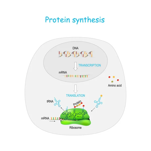
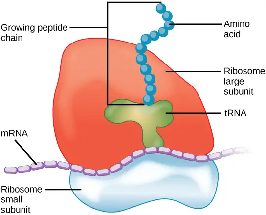
Ribosomes vector illustration - VectorMine Most Vector Editing Software. 3. High-resolution JPG image. 3800 x 3965 px. License terms in short: Use for everything except reselling item itself. Read a full license here. Description: Ribosomes vector illustration. Anatomical and medical labeled scheme with tRNA, Amino acid, protein, cell, small and large subunit, mRNA.
A Labeled Diagram of the Animal Cell and its Organelles A Labeled Diagram of the Animal Cell and its Organelles There are two types of cells - Prokaryotic and Eucaryotic. Eukaryotic cells are larger, more complex, and have evolved more recently than prokaryotes. Where, prokaryotes are just bacteria and archaea, eukaryotes are literally everything else.
Biuret test - Wikipedia The Biuret (IPA: / ˌ b aɪ j ə ˈ r ɛ t /, / ˈ b aɪ j ə ˌ r ɛ t /) test, also known as Piotrowski's test, is a chemical test used for detecting the presence of peptide bonds (Note that at least two peptide bonds are needed to be present in the molecule to show this test). In the presence of peptides, a copper(II) ion forms mauve-colored coordination complexes in an alkaline solution.
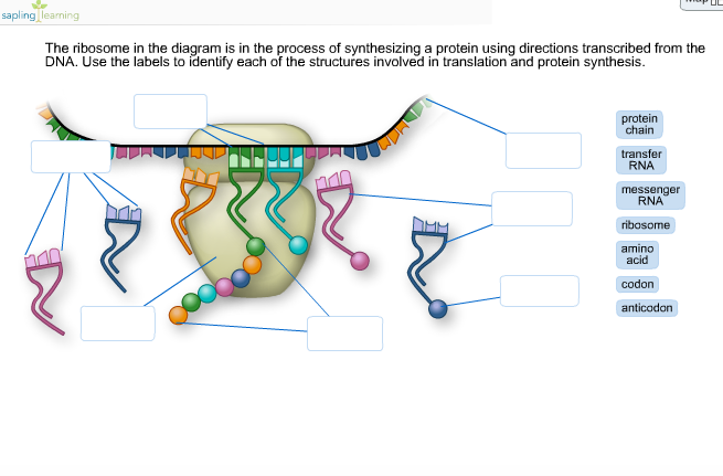
Ribosome and protein synthesis, diagram - Stock Image - C029/3020 Diagram showing protein synthesis in cells (translation). Messenger ribonucleic acid (mRNA, blue with coloured nucleotides) is read by a ribosome (pink). The molecules of transfer RNA (tRNA, key-shaped) each bring an amino acid (orange dot) to bind to the ribosome's protein synthesis site.
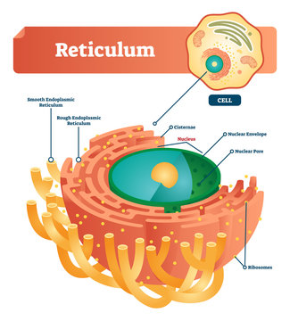
Nuclear envelope - Wikipedia The nuclear envelope is punctured by around a thousand nuclear pore complexes, about 100 nm across, with an inner channel about 40 nm wide. The complexes contain a number of nucleoporins, proteins that link the inner and outer nuclear membranes.. Cell division. During the G2 phase of interphase, the nuclear membrane increases its surface area and doubles its …
Active Ribosome Profiling with RiboLace - PubMed Ribosome profiling, or Ribo-seq, is based on large-scale sequencing of RNA fragments protected from nuclease digestion by ribosomes. Thanks to its unique ability to provide positional information about ribosomes flowing along transcripts, this method can be used to shed light on mechanistic aspects …
Parts of a Mitochondria Diagram | Ribosomes & Function of Mitochondrial ... Ribosomes: Mitochondrial ribosomes help to translate various proteins in the mitochondrial matrix Metabolites : Mitochondria contain metabolites that are involved in the citric acid cycle.
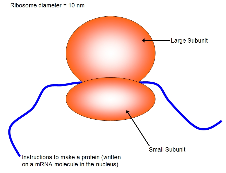
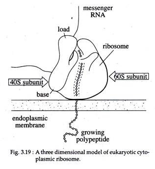
Ribosomes: Structure, Composition, and Assembly (With Diagram) Ribosomes in the cytoplasm of eukaryotic cells have a sedimentation coefficient of about 80 S (MW about 4.5 x 10 6) and are composed of 40 S and 60 S subunits. In prokaryotic cells, ribosomes are typically about 70 S (MW about 2.7 x 10 6) and are formed from 30 S and 50 S subunits.
› questions-and-answers › a-what-isAnswered: a) What is a mutation in molecular… | bartleby For example, part of the viral PA gene includes a rarely used CGU codon. When the ribosome pauses to translate this codon, it may slip ahead by one nucleotide and produce a polypeptide with a diff erent C-terminal sequence. From the partial mRNA sequence shown here, determine the normal polypeptide sequence and the sequence with the frameshift.
GRADE 12 LIFE SCIENCES LEARNER NOTES - Mail & Guardian 1.1 The diagram below represents a part of a molecule. Study the diagram and answer the questions that follow. 1.1.1 Identify the molecule in the above diagram. (1) 1.1.2 Label the parts numbered 1 and 5 respectively. (2) 1.1.3 What is the collective name …
serve.mg.co.za › content › documentsGRADE 12 LIFE SCIENCES LEARNER NOTES - Mail & Guardian 1.1 The diagram below represents a part of a molecule. Study the diagram and answer the questions that follow. 1.1.1 Identify the molecule in the above diagram. (1) 1.1.2 Label the parts numbered 1 and 5 respectively. (2) 1.1.3 What is the collective name for the parts numbered 2, 3 and 4? (1)
› articles › s41586/021/04204-9ER proteins decipher the tubulin code to regulate organelle ... Dec 15, 2021 · Since full-length p180 (p180L) is degraded during cell lysis (Extended Data Fig. 3a, c), we used a smaller, more stable splice variant (p180s) that lacks the numerous ribosome-binding decapeptide ...
Solved The ribosome in the diagram is in the process of | Chegg.com The ribosome in the diagram is in the process of synthesizing a protein using directions transcribed from the DNA. Use the labels to identify each of the structures involved in translation and protein synthesis. Question: The ribosome in the diagram is in the process of synthesizing a protein using directions transcribed from the DNA.
GENOMICS - molbiol-tools.ca In programmed frameshifting, the ribosome switches to an alternative frame at a specific site in response to a special signal in a messanger RNA. Programmed frameshift plays role in viral particle morphogenesis, autogenous control, and alternative enzymatic activities. The common frameshift is a -1 frameshift, in which the ribosome shifts a single nucleotide in the upstream …
Ribosomes Vector Illustration. Anatomical and Medical Labeled Scheme ... Ribosomes vector illustration. Anatomical and medical labeled scheme. Explained closeup diagram.. Illustration about amino, educational, biogenesis, labeled, golgi, body - 122097933
protein synthesis diagram labeled - TheFitnessManual Switch RNAs (tRNAs) deliver amino acids to the ribosome. - "protein synthesis diagram labeled" tRNAs are additionally RNA polymers. They're typically between 75 and 90 RNA nucleotides lengthy. However in contrast to mRNAs, that are linear, hydrogen bonding between nucleotides inside a tRNA causes it to fold up.
molbiol-tools.ca › GenomicsGENOMICS - molbiol-tools.ca These multivariate analyses can be done using either taxonomic or automatically generated phenotypic labels and visualized using a variety of high quality graphical tools. The bacterial census data can be derived from 16S rRNA data, NextGen shotgun sequencing or even classical microbial culturing techniques. Includes a tutorial.
Molecular Biology, Robert Weaver, 5th Edition - Academia.edu Canadian journal of gastroenterology = Journal canadien de gastroenterologie. Applications of recombinant DNA technology in gastrointestinal medicine and hepatology: Basic paradigms of molecular cell biology.
Bio101 - Ch 6 HW Flashcards | Quizlet Tour of an Animal Cell: Part A. Drag the labels on the left onto the diagram of the animal cell to correctly identify the function performed by each cellular structure. a. smooth ER- synthesizes lipids. b. nucleolus- assembles ribosomes. c. defines cell shape.
Animal cell diagram to label - Labelled diagram Drag and drop the pins to their correct place on the image.. ribosome, nucleus, cytoplasm, cell membrane, mitochondria. ribosome, nucleus, cytoplasm, cell membrane, mitochondria. Animal cell diagram to label
Looking at the Structure of Cells in the Microscope A typical animal cell is 10–20 μm in diameter, which is about one-fifth the size of the smallest particle visible to the naked eye. It was not until good light microscopes became available in the early part of the nineteenth century that all plant and animal tissues were discovered to be aggregates of individual cells. This discovery, proposed as the cell doctrine by Schleiden and …
Structure of Ribosome - Biology Wise Diameter of Ribosome is 20nm. Their composition can be divided into two parts - 2/3 part of r-RNA (ribosomal RNA) and 1/3 part RNP (Ribosomal protein or Ribonuclep protein). Polypeptide chain is fabricated by translating mRNA (messenger RNA) with the aid amino acids that tRNA (transfer RNA) delivers.
Structure of Ribosome (With Diagram) - Biology Discussion A bacterial ribosome is about 250 nm in diameter and consists of two subunits, one large and one small. Both subunits consist of one or more molecules of rRNA and an array of ribosomal proteins. ADVERTISEMENTS: Association of two subunits is called mono-some. The structure of prokaryotic ribosome is given in the figure 8.2 B.
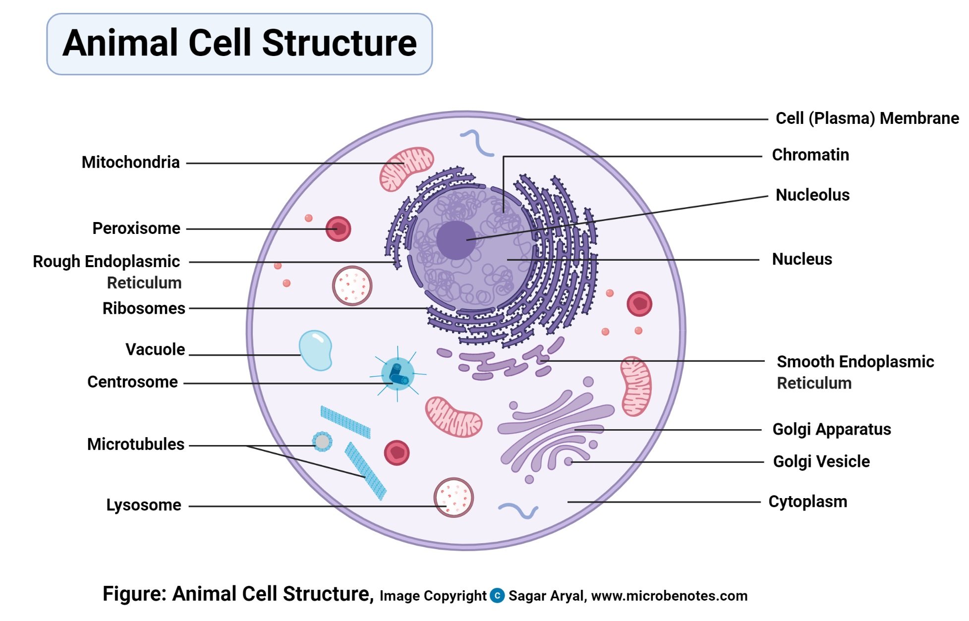
Animal Cell Diagram | Science Trends The diagram, like the one above, will include labels of the major parts of an animal cell including the cell membrane, nucleus, ribosomes, mitochondria, vesicles, and cytosol. The cells of animals are the basic structural units for the wide variety of life we see in the animal kingdom.
Ribosome - Definition, Function and Structure | Biology Dictionary A. Ribosomes translate the 4 base language of DNA into the 20 base language of proteins, allowing for many more combinations. B. The 4 different nucleobases of DNA can be recombined endlessly to produce new proteins. C. Ribosomes can modify proteins with carbohydrates to make them unique. Answer to Question #2 3.















Post a Comment for "41 ribosome diagram with labels"