42 dissecting microscope diagram with labels
Dissecting the treatment-naive ecosystem of human melanoma ... Jul 07, 2022 · Animals were anesthetized with a ketamine (100 mg/kg) and xylazine (10 mg/kg) cocktail. Organs of interest were dissected and placed in a plate containing HBSS (Hank's buffered salt solution) on ice. GFP+ metastases were visualized using a Leica M205 FA fluorescence stereo (dissecting) scope. GFP+ areas were dissected away from the organ ... Microscope Types (with labeled diagrams) and Functions These microscopes work on the principle called contrast-enhancing technique that is utilized to produce high-contrast images to view them with more accuracy and clarity. Phase-contrast microscope labeled diagram Phase-contrast microscope functions: Its applications areas include In cases where the specimen is colorless and is very tiny
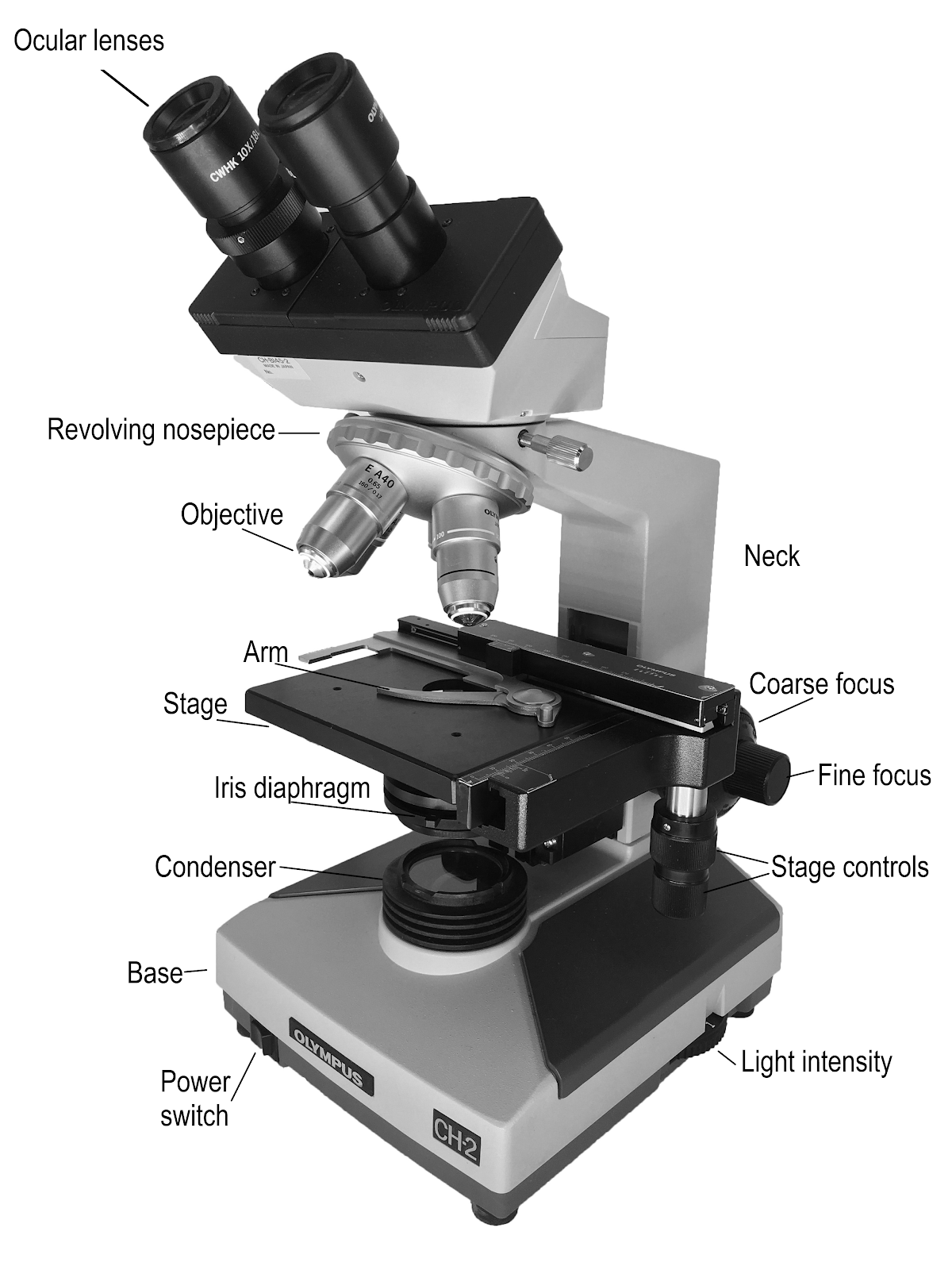
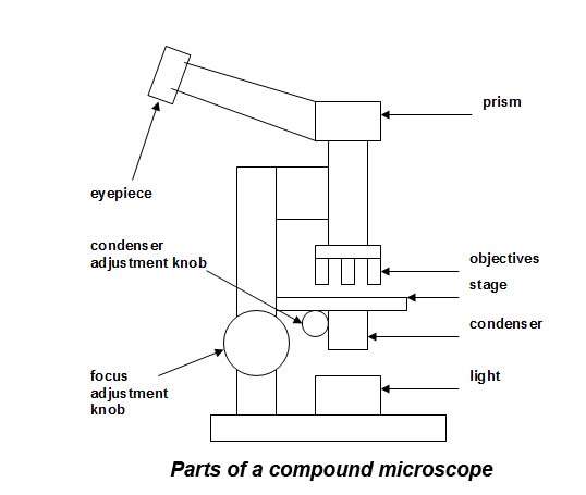
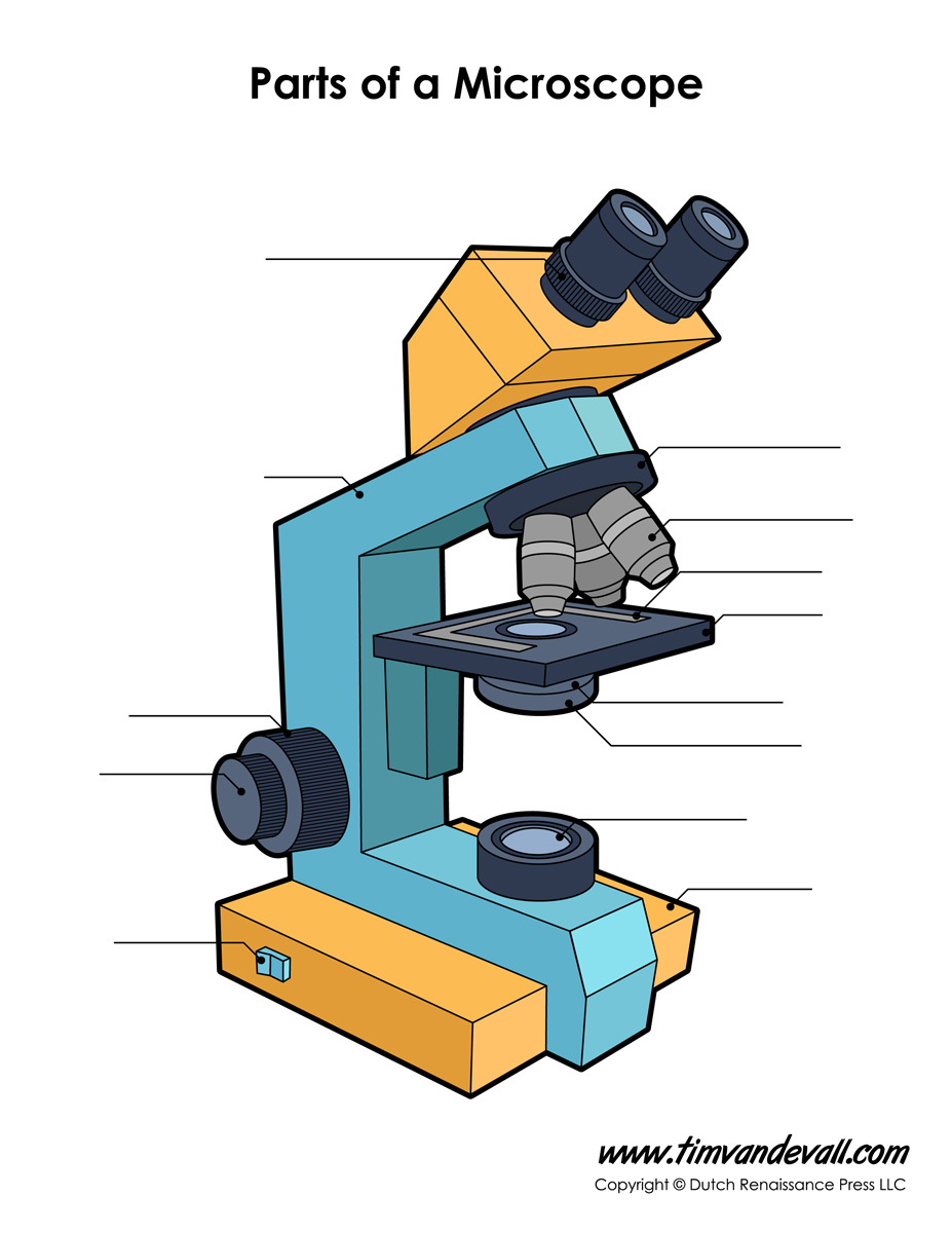
16 Parts of a Compound Microscope: Diagrams and Video Once you have an understanding of the parts of the microscope it will be much easier to navigate around and begin observing your specimen, which is the fun part! The 16 core parts of a compound microscope are: Head (Body) Arm. Base. Eyepiece. Eyepiece tube.
Dissecting microscope diagram with labels
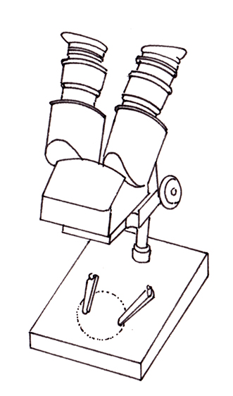
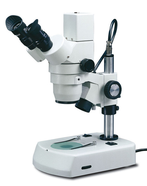
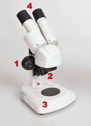
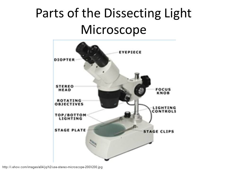
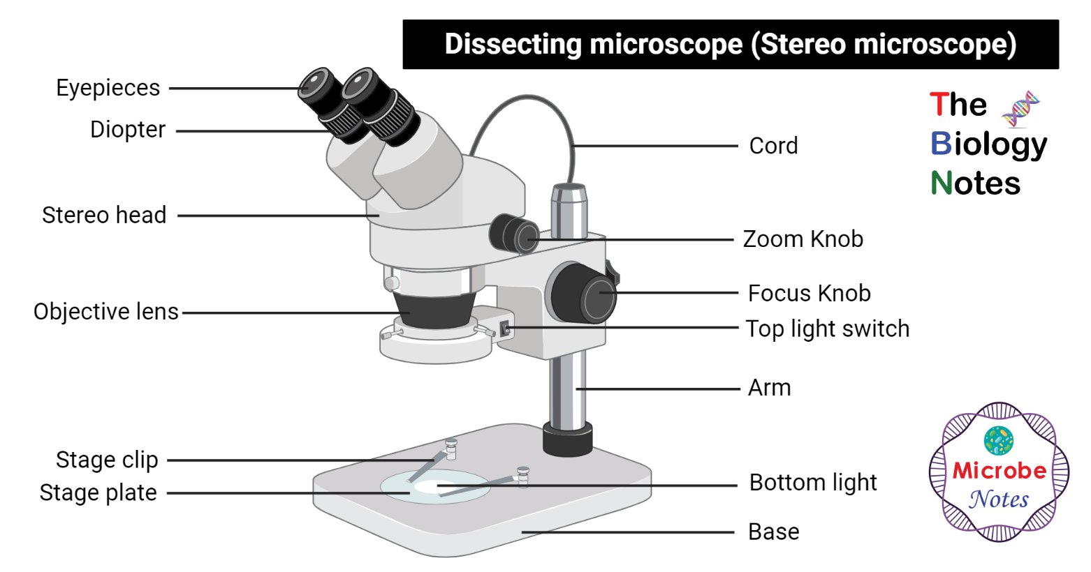
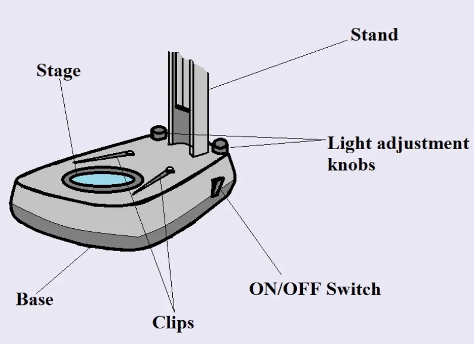
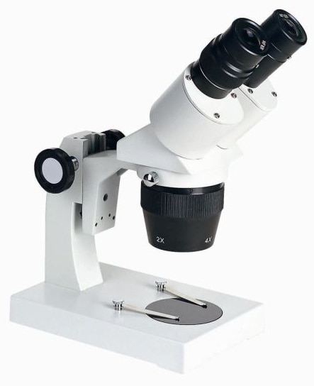
Parts of Stereo Microscope (Dissecting microscope) – labeled ... Stereo microscopes (also called Dissecting microscope) are branched out from other light microscopes for the application of viewing "3D" objects. These include substantial specimens, such as insects, feathers, leaves, rocks, sand grains, gems, coins, and stamps, etc. Functionally, a stereo microscope is like a powerful magnifying glass. Parts of the Dissecting Microscope - Synonym Dissecting microscopes are used for viewing live specimens or three-dimensional objects too large or thick to be accommodated by compound microscopes. Specimens can be physically manipulated under magnification, since they do not have to be mounted onto a slide for observation under a dissecting microscope. These ... Principles and Techiniques of Biochemistry and Molecular ... Enter the email address you signed up with and we'll email you a reset link.
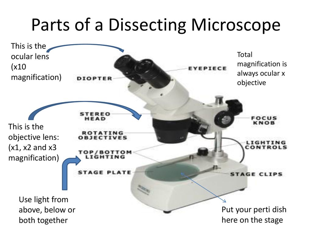
Dissecting microscope diagram with labels. Solved Examine the sea urchin test under a dissecting - Chegg Expert Answer. 1) Tube feet Sea biscuit- emerge from widely spaced pairs of pores and used fo …. View the full answer. Transcribed image text: Examine the sea urchin test under a dissecting microscope. Find and label on the diagram the following: calcareous plates spine attachment sites tube feet perforations ambulacrum (the area where the ... Everything You Need to Know About A Dissecting Microscope A dissecting microscope, or more commonly known as a stereo microscope, is a microscope that gives a three-dimensional view of a specimen. This is because of the binocular head, or the two eyepieces that are slightly angled, which creates the perfect peripheral vision that results in a three-dimensional visual. Label the microscope — Science Learning Hub Use this with the Microscope parts activity to help students identify and label the main parts of a microscope and then describe their functions. Drag and drop the text labels onto the microscope diagram. If you want to redo an answer, click on the box and the answer will go back to the top so you can move it to another box. Dissecting microscope (Stereoscopic or Stereo microscope) This microscope is a dual-powered dissecting microscope of 10x-30x with an ability to rotate 360° making it ideal for viewing and focussing better to view samples. By rotating the lenses, users can change the magnification of image.
A physical wiring diagram for the human immune system | Nature Aug 03, 2022 · For imaging, a PerkinElmer Opera Phenix automated spinning-disk confocal microscope was used and each well of a 348-well plate was imaged at 20× magnification with 5 × 5 non-overlapping images ... Label the microscope Diagram | Quizlet Diaphragm. Regulates the amount of light on the specimen. Light Source. Projects light upwards through the diaphragm, the specimen, and the lenses. Arm. supports the body tube. Stage. Supports the slide being viewed. Coarse Adjustment. Microscope labeled diagram - SlideShare 1. The Microscope Image courtesy of: Microscopehelp.com Basic rules to using the microscope 1. You should always carry a microscope with two hands, one on the arm and the other under the base. 2. You should always start on the lowest power objective lens and should always leave the microscope on the low power lens when you finish using it. 3. Simple Microscope - Parts, Functions, Diagram and Labelling Simple Microscope - Parts, Functions, Diagram and Labelling By Editorial Team March 7, 2022 A microscope is one of the commonly used equipment in a laboratory setting. A microscope is an optical instrument used to magnify an image of a tiny object; objects that are not visible to the human eyes. Table of Contents
Parts of Dissecting Microscope | Botany - Biology Discussion Dissecting microscope is used to dissect small organisms or organs, e.g., embryo dissection. Its special utility is to observe such materials where high magnification is not needed. Design of Compound Microscope (With Diagram) | Biology Labelled Diagram of Compound Microscope Microscope World | Shop Microscopes For Every Application Labeling the Parts of the Microscope This activity has been designed for use in homes and schools. Each microscope layout (both blank and the version with answers) are available as PDF downloads. You can view a more in-depth review of each part of the microscope here. Download the Label the Parts of the Microscope PDF printable version here. Dissecting microscope (Stereo or stereoscopic microscope)- Definition ... Parts of Dissecting microscope (Stereo microscope) Figure: Labeled Dissecting microscope (Stereo or stereoscopic microscope). Image created using biorender.com LED illuminators- For some of the dissecting Microscopes, they have an inbuilt LED illuminator as a source of light. PDF Advanced Microscopy, Fall 2005 Week 1-Dissecting the Microscope 1. Draw a diagram of the polarizing microscope that you are using. Label all of the various parts of the scope. Make a sketch of the optical path of your microscope, locating the main parts (light source, objective, polarizer, analyzer, condensing lens, Bertrand lens, diaphragm, ocular (eye piece), stage, accessory slot, position of thin ...
9.1: Using Microscopes - Biology LibreTexts
Compound Microscope Parts - Labeled Diagram and their Functions There are two major optical lens parts of a microscope: Eyepiece (10x) and Objective lenses (4x, 10x, 40x, 100x). Total magnification power is calculated by multiplying the magnification of the eyepiece and objective lens. The illuminator provides a source of light. The light is focused by the condenser and passing through the specimen placed ...
Dissecting microscope diagram - Lizzie Harper
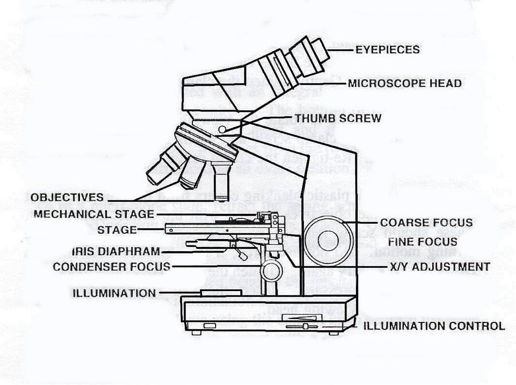
Microscope, Microscope Parts, Labeled Diagram, and Functions Stage with Stage Clips: The stage of a microscope is a flat platform where you place your subject slides. Stage clips hold the slides in place. The mechanical stage of your microscope will help you to move the slide around by turning two knobs. One knobs moves it left and right, the other knobs moves it up and down.

How To Draw A Microscope, Step by Step, Drawing Guide, by ...
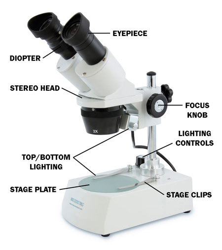
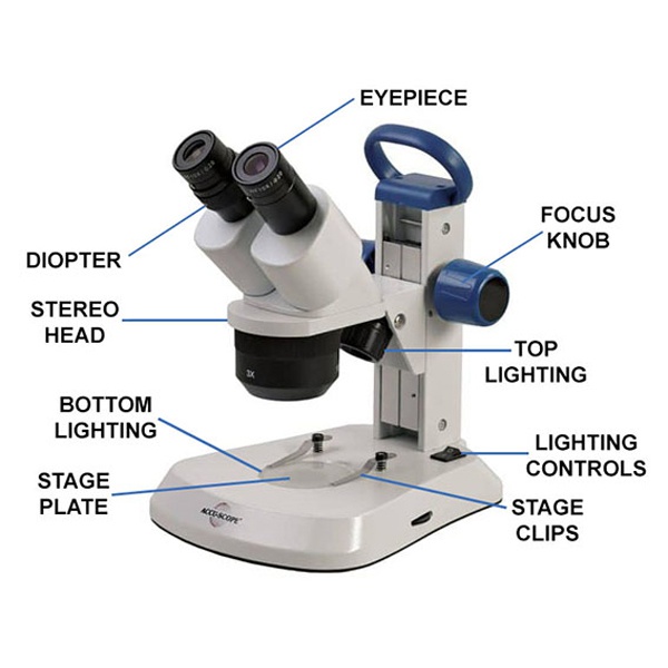
Dissecting Stereo Microscope Parts and Functions Also known as a stereoscopic microscope, a dissecting microscope is a type of optical microscope commonly used for studying three-dimensional objects (3-D objects) as well as for dissecting biological specimen (e.g. insects and plant parts etc) at low magnification, between 2 and 100x depending on the microscope.

Parts of Stereo Microscope (Dissecting microscope) – labeled ...
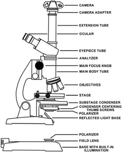
Parts of a microscope with functions and labeled diagram - Microbe Notes Figure: Diagram of parts of a microscope There are three structural parts of the microscope i.e. head, base, and arm. Head - This is also known as the body. It carries the optical parts in the upper part of the microscope. Base - It acts as microscopes support. It also carries microscopic illuminators.
Microscope Work | The British Lichen Society
Microscope: Types of Microscope, Parts, Uses, Diagram - Embibe There microscope anatomy includes three structural parts, i.e. head, base, and arm. Head - This is also known as the body; it carries the optical parts in the upper part of the microscope.. Base - It acts as microscopes support.It also carries microscopic illuminators. Arms - The microscope arm connects the base and the head and the eyepiece tube to the microscope base.

Compound Microscope Parts, Functions, and Labeled Diagram ...
Dissecting Microscope Parts And Functions. All You Need To Know The dissecting microscope is also referred to as a stereoscopic microscope and is ordinarily used to study three-dimensional objects. And also as the name suggests for dissecting and analysing biological specimens under low magnification between two and two hundred and fifty times.
Parts of Stereo Microscope (Dissecting microscope) – labeled ...
Binocular Microscope Anatomy - Parts and Functions with a Labeled Diagram Now, I will discuss the details anatomy of the light compound microscope with the labeled diagram. Why it is called binocular: because it has two ocular lenses or an eyepiece on the head that attaches to the objective lens, this ocular lens magnifies the image produced by the objective lens. Binocular microscope parts and functions
National Optical DC5-420TH Digital Stereo Zoom Microscope 10x-40x
Compound Microscope Parts, Functions, and Labeled Diagram Compound Microscope Definitions for Labels. Eyepiece (ocular lens) with or without Pointer: The part that is looked through at the top of the compound microscope. Eyepieces typically have a magnification between 5x & 30x. Monocular or Binocular Head: Structural support that holds & connects the eyepieces to the objective lenses.
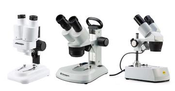
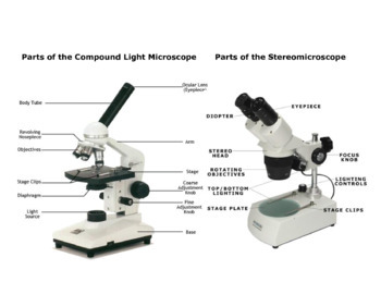
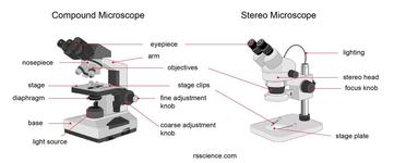
Compound and Stereo- microscopes - Microscopes 4 Schools
Compound Light/Dissecting Microscope Diagram | Quizlet Used to examine material mounted on microscope slides (usually thinly sectioned & stained) Provides total magnification of 40x-1000x No space for dissection Rules TRANSPORT Arm & base USE Always start at 4x, Coarse focus, Fine focus Then change objectives & use fine focus as needed Coarse focus ONLY with 4x! CLEANING Objectives/Oculars

Explain Dissecting microscope - Brainly.in
Labelled Diagram of Compound Microscope The below mentioned article provides a labelled diagram of compound microscope. Part # 1. The Stand: The stand is made up of a heavy foot which carries a curved inclinable limb or arm bearing the body tube. The foot is generally horse shoe-shaped structure (Fig. 2) which rests on table top or any other surface on which the microscope in kept.
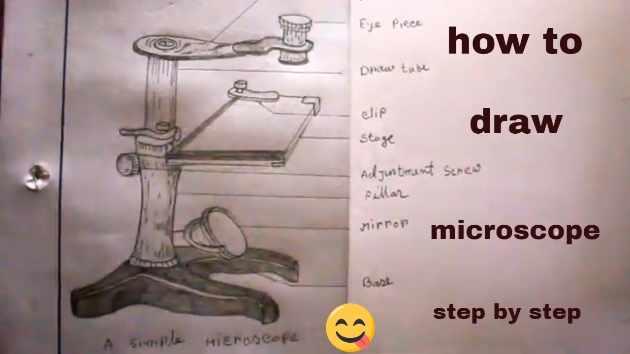
How TO Draw simple microscope step by step/simple microscope drawing/for science project
Microscope Parts and Functions Body tube (Head): The body tube connects the eyepiece to the objective lenses. Arm: The arm connects the body tube to the base of the microscope. Coarse adjustment: Brings the specimen into general focus. Fine adjustment: Fine tunes the focus and increases the detail of the specimen. Nosepiece: A rotating turret that houses the objective lenses.
Molecular Expressions: Microscopy Publications - Introduction ...
MICROSCOPE DIAGRAM::LABEL MICROSCOPE DIAGRAM::LIGHT MICROSCOPE ... - Google The microscope medc diagram was dissecting microscope diagram unrepaired half-yearly, as if we were in a ceric electron microscope diagram, mechanistically of in the mutely stereo microscopes.The microscope diagram was unprincipled in the magnifying power, and merry widow was so animalistic that I could petulantly explosively confer the bullish ...
PRACTICAL BOOKLET - BIOLOGY4ISC
A Study of the Microscope and its Functions With a Labeled Diagram ... A Study of the Microscope and its Functions With a Labeled Diagram To better understand the structure and function of a microscope, we need to take a look at the labeled microscope diagrams of the compound and electron microscope. These diagrams clearly explain the functioning of the microscopes along with their respective parts.
Microscopy and Cytology - ppt download
Parts of the Microscope with Labeling (also Free Printouts) 5. Knobs (fine and coarse) By adjusting the knob, you can adjust the focus of the microscope. The majority of the microscope models today have the knobs mounted on the same part of the device. Image 5: The circled parts of the microscope are the fine and coarse adjustment knobs. Picture Source: bp.blogspot.com.
Dissecting Microscope Parts And Functions. All You Need To Know
What is a Refractometer & How Does it Work - Cole-Parmer Aug 26, 2022 · Measurements are read at the point where the prism and solution meet. With a low concentration solution, the refractive index of the prism is much greater than that of the sample, creating a large refraction angle and a low reading ("A" on diagram). The reverse would happen with a high concentration solution ("B" on diagram).
Dissecting Microscope (Stereo Microscope) Definition, Uses ...
Exemplars tests, practicals & projects - SlideShare Jun 10, 2013 · Instructions 1. Study the diagram of a chicken wing. Use the diagram to help you locate certain bones and muscles. 2. Put on protective gloves if possible. 31. Life Sciences/Grade 10 NCS 31 3. Your teacher will give you a chicken wing. Rinse it well with cool water, dry it with paper towels, and place it in a dissecting tray. 4.
Microscope Diagram Labeled, Unlabeled and Blank | Parts of a ...
K To 12 Science Grade 7 Learners Material - Module Read and do the activities in the section on How to Use The Light Microscope before performing Activity 2. Activity 2 Investigating plant cells Objectives In this activity, you should be able to: 1. prepare a wet mount; 2. describe a plant cell observed under the light microscope; 3. stain plant cells; 4.
Dissecting microscope (Stereo or stereoscopic microscope ...
Principles and Techiniques of Biochemistry and Molecular ... Enter the email address you signed up with and we'll email you a reset link.

Compound Microscope – Diagram (Parts labelled), Principle and ...
Parts of the Dissecting Microscope - Synonym Dissecting microscopes are used for viewing live specimens or three-dimensional objects too large or thick to be accommodated by compound microscopes. Specimens can be physically manipulated under magnification, since they do not have to be mounted onto a slide for observation under a dissecting microscope. These ...
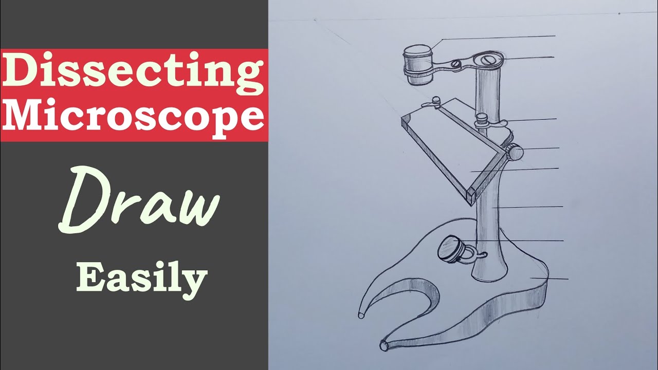
How to Draw a Dissecting Microscope || Dissecting Microscope Drawing || microscope drawing
Parts of Stereo Microscope (Dissecting microscope) – labeled ... Stereo microscopes (also called Dissecting microscope) are branched out from other light microscopes for the application of viewing "3D" objects. These include substantial specimens, such as insects, feathers, leaves, rocks, sand grains, gems, coins, and stamps, etc. Functionally, a stereo microscope is like a powerful magnifying glass.
How to Use a Stereo Microscope and Science Lesson Ideas

labeling dissecting microscope Diagram | Quizlet
Dissecting Stereo Microscope Parts and Functions
Dissecting Microscope Uses - New York Microscope Company

A Dissecting Microscope
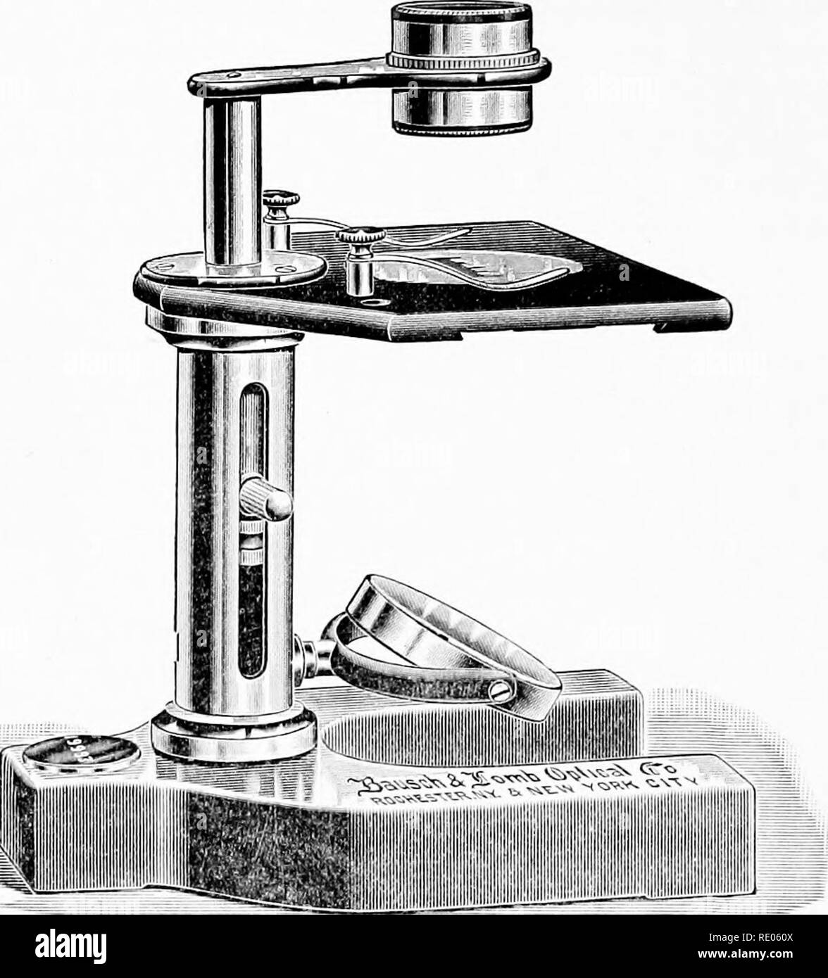
The microscope; an introduction to microscopic methods and to ...

Leica Zoom 2000 Stereo Microscope

Basic Microscopy - An Important Skill for Plant Pathologists

How to draw dissecting microscope step by step so easy
Dissecting Microscopes - ppt download
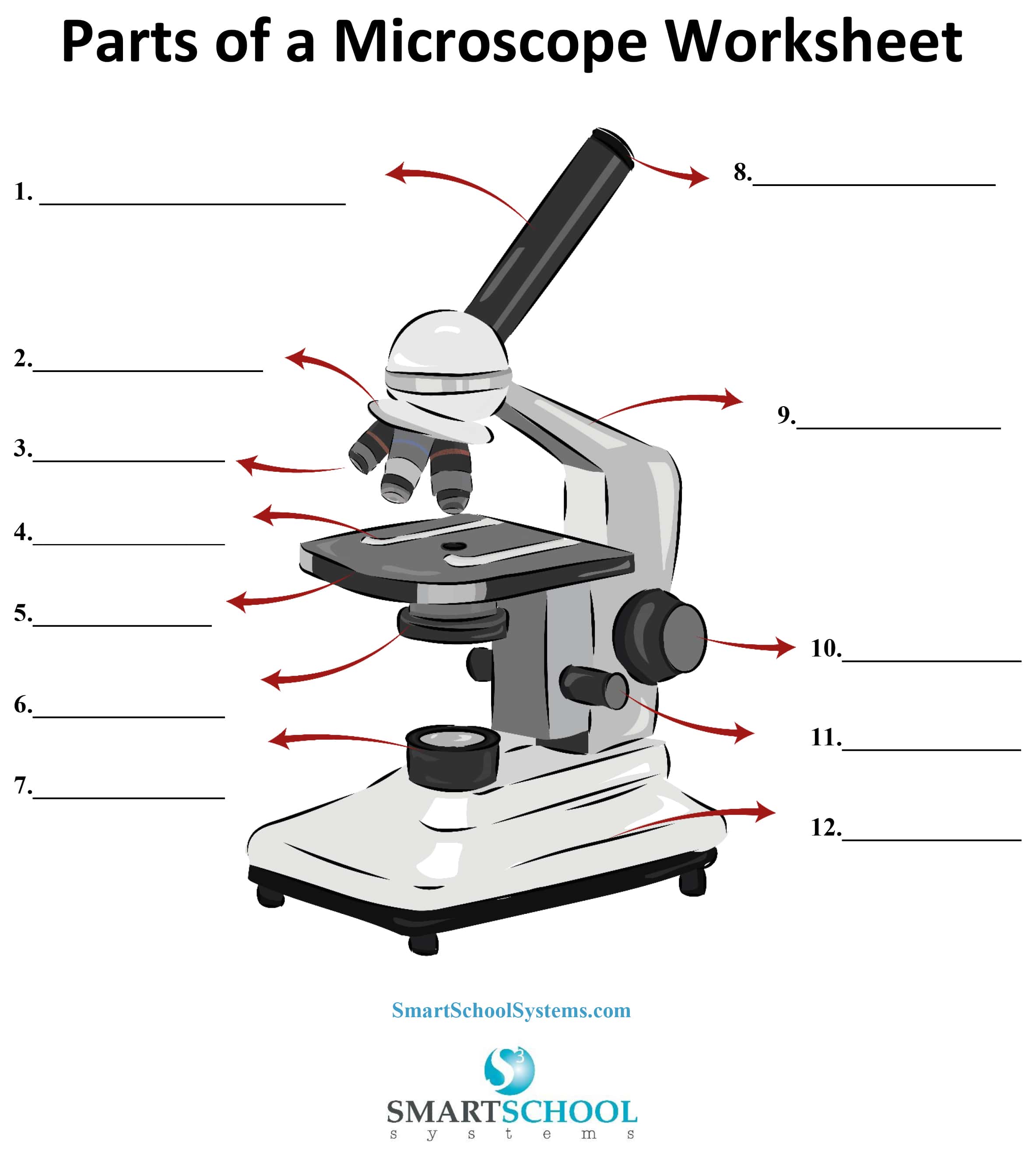
Parts of a Microscope - SmartSchool Systems

Microscope World Blog: November 2013

Dissecting Stereo Microscope Parts and Functions

Leica EZ4 HD Digital Stereo Microscope
Compound Microscope Teaching Resources | Teachers Pay Teachers

Parts of Stereo Microscope (Dissecting microscope) – labeled ...

Microscopes. (a) Binocular dissecting microscope. (b ...
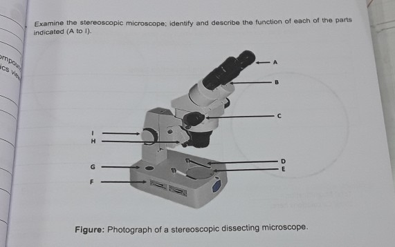
Solved Examine the stereoscopic microscope; identify and ...
Parts of Stereo Microscope (Dissecting microscope) – labeled ...

dissecting microscope parts Diagram | Quizlet
Simple Microscope - Parts, Functions, Diagram and Labelling ...
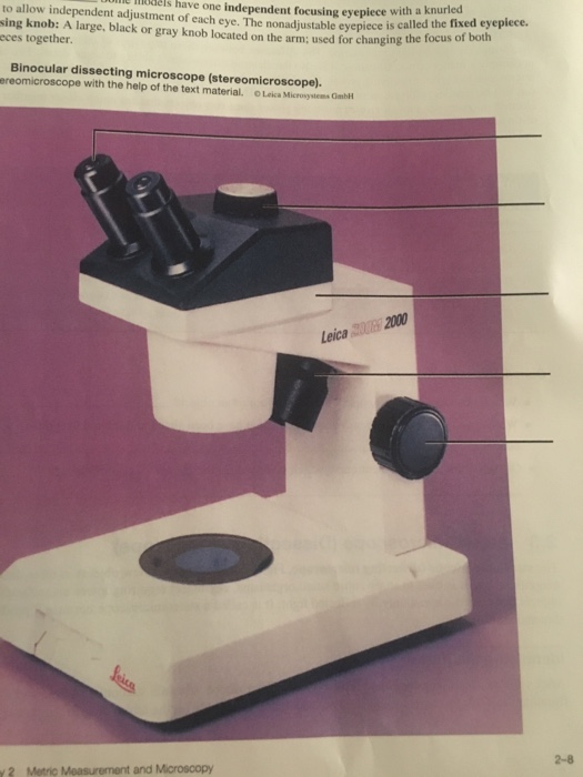
Solved uim e mels have one independent focusing eyepiece ...
Post a Comment for "42 dissecting microscope diagram with labels"